Operation of the CEM Discover SP Microwave Reaction System for Amino Acid Hydrolysis
5.1 INTRODUCTION
As noted earlier, various equipment exists specifically to assist in the hydrolysis of samples. Regardless of the instrumentation, there are usually three different hydrolysis protocols: acid hydrolysis, used to determine the total protein composition; acid hydrolysis following performic acid oxidation, required to measure sulfur-containing amino acids such as cysteine and methionine; and alkaline hydrolysis, used to assess tryptophan recovery. While traditional equipment for sample hydrolysis often involves a time-consuming process, instrumentation for microwave hydrolysis can significantly speed up things. Implementation of microwave hydrolysis for all three protocols results in improved control of hydrolysis conditions with better accuracy, reproducibility, speed, and robustness.
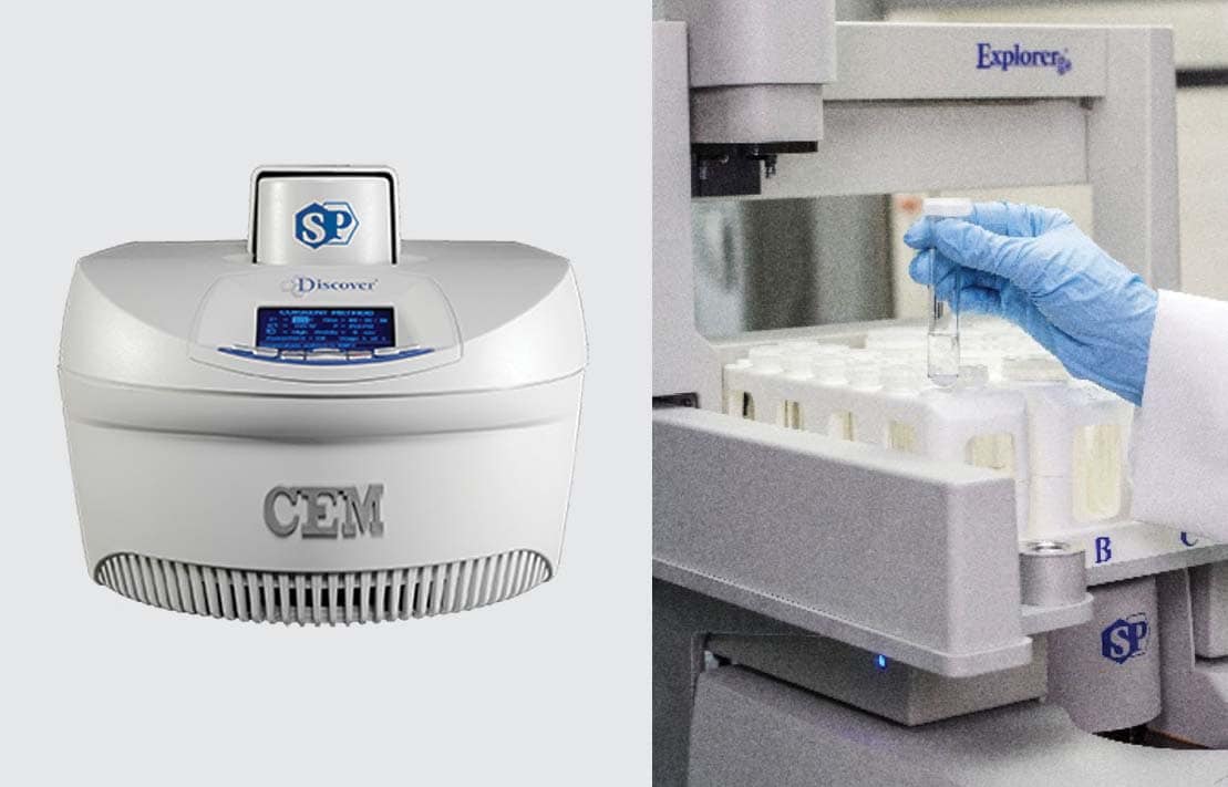
Figure 7. CEM Discover SP Microwave Reaction System with Explorer Automation.
Using microwave technology, the CEM Discover SP Microwave Reaction System (CEM Corporation 3100 Smith Farm Road Matthews, NC 28104) (Figure 7) can hydrolyze proteins and peptides in preparation for amino acid analysis in just 15 minutes. As illustrated in Figure 8, direct application of microwave energy and use of integrated IR temperature sensors allow for rapid heating and maintenance of the set temperature for each sample. In situ stirring ensures even heat distribution through the sample, and post-reaction quenching cools the sample in seconds.
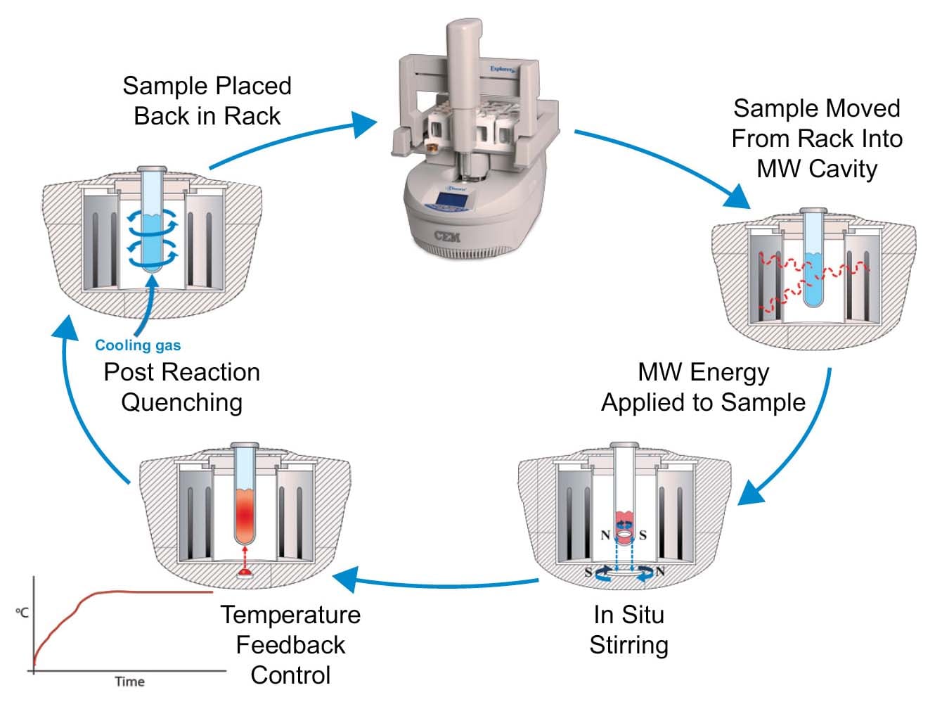
Figure 8. Process of microwave hydrolysis in the CEM Discover SP.
The following section outlines system setup and operation. For further information, contact CEM Corporation or refer to the system manual.
5.2 PHYSICAL REQUIREMENTS
- The system has an electrical requirement of 110–140 VAC, 60 Hz, 10 A at 120 VAC.
- The system requires venting.
- The system requires a minimum of 25 psi and a maximum of 60 psi compressed air or compressed nitrogen.
- The system requires a laptop (provided by CEM) located within ten feet of the system.
5.3 REAGENTS REQUIREMENTS
- 6 N HCl for acid hydrolysis, 1:1 concentrated HCl:DI water.
- 4 N NaOH for base hydrolysis, dissolve 16 g of solid NaOH in 100 mL of DI water.
- Performic acid (preoxidation only), 9:1 formic acid:30% hydrogen peroxide.
- 10 or 35 mL vial for Discover SP with stir bar and cap.
- Phenol crystals.
5.4 SAMPLE PREPARATION FOR ACID OR BASE HYDROLYSIS
Preoxidation is required to analyze cysteine and methionine. Base hydrolysis is required to analyze tryptophan. It is not recommended that you run more than a 500-mg sample in a 35-mL vial.
5.4.1 Performic acid oxidation (preoxidation)
Option 1
- Weigh 50–500 mg of sample into a 35-mL vial with stir bar.
- Place digestion tubes in an ice bath for about 15 minutes.
- After cooling, add 5 mL of performic acid.
- Cover the tubes with glass stoppers and stir on a magnetic stirring plate at low speed for 15 minutes.
- Return the digestion tubes to the ice bath and let oxidize for 16 hours.
- Remove the glass stoppers and add 0.84 g of sodium metabisulfite to decompose performic acid.
- Perform acid hydrolysis starting on Step 2 of Option 1 in section 5.4.2 Acid Hydrolysis.
Option 2
- Weigh 50–500 mg of sample into a 35-mL vial with stir bar.
- Add 6 mL of freshly prepared performic acid solution (9:1 formic acid:30% hydrogen peroxide).
- Incubate the reaction mix in a water bath at 50 °C for one hour.
- Evaporate residual performic acid solution to dryness in an evaporator at 50 °C.
- Perform acid hydrolysis starting on Step 2 of Option 1 in section 5.4.2 Acid Hydrolysis.
5.4.2 Acid hydrolysis
Option 1: 50–300 mg sample
- Weigh the sample into a 35-mL vial with stir bar.
- Add 3% by weight of phenol.
- Add 5 mL of 6 N HCl.
- Cap the vial.
- Using Dynamic Method Control, program a method with 300 W, 15-minute hold, 195 °C, and 300 psi. Stirring should be set to high.
- Place the vial into the Discover Cavity and press the Start Key to begin the method.
- The sample should heat to 195 °C, at no more than 250 psi (±10%).
Option 2: 300–500 mg sample
- Weigh the sample into a 35-mL vial with stir bar.
- Add 3% by weight of phenol.
- Add 10 mL of 6 N HCl.
- Cap the vial.
- Using Dynamic Method Control, program a method with 300 W, 15-minute hold, 195 °C, and 300 psi. Stirring should be set to high.
- Place the vial into the Discover Cavity and press the Start Key to begin the method.
- The sample should heat to 195 °C, at no more than 250 psi (±10%).
5.4.3 Base hydrolysis
Option 1: 50–300 mg sample
- Weigh the sample into a 35-mL vial with stir bar.
- Add 5 mL of 4 N NaOH.
- Cap the vial.
- Using Dynamic Method Control, program a method with 300 W, 15-minute hold, 195 °C, and 300 psi. Stirring should be set to high.
- Place the vial into the Discover Cavity and press the Start Key to begin the method.
- The sample should heat to 195 °C, at no more than 250 psi (±10%).
Option 2: 300–500 mg sample
- Weigh the sample into a 35-mL vial with stir bar.
- Add 10 mL of 4 N NaOH.
- Cap the vial.
- Using Dynamic Method Control, program a method with 300 W, 15-minute hold, 195 °C, and 300 psi. Stirring should be set to high.
- Place the vial into the Discover Cavity and press the Start Key to begin the method.
- The sample should heat to 195 °C, at no more than 250 psi (±10%).
5.5 RECOMMENDATIONS FOR CEM MICROWAVE HYDROLYSIS
The temperature, time and power settings are dependent of the types of samples being hydrolyzed. It's important to maintain the microwave to be functioning all of the way through the procedure. This can require some experimentation by the user to best optimize their samples using this technique.
It is recommended that you use the correct sample size, volume, and vessels in the procedures described earlier. These factors are critical to the success of the hydrolysis. Some components of the sample will remain in the solid phase after hydrolysis, and they typically turn black during the process, as shown in Figure 9. This occurrence is normal; the amino acids will be in solution. Please direct all questions related to use of CEM equipment to CEM Corporation via molecular.support@cem.com or by calling 1-704 -821-7015 and requesting the CEM Molecular Sample Preparation Team.
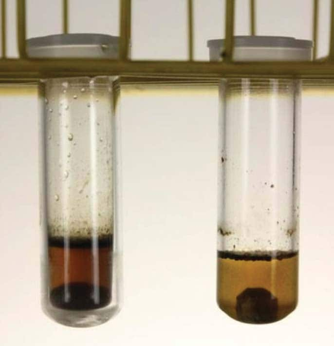
Figure 9. A hydrolyzed sample.